Figure 1. [The normal human retina fundus]. - Webvision - NCBI
Por um escritor misterioso
Descrição
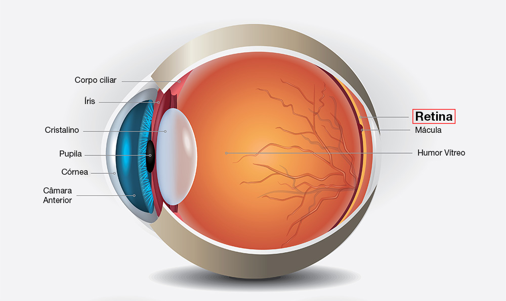
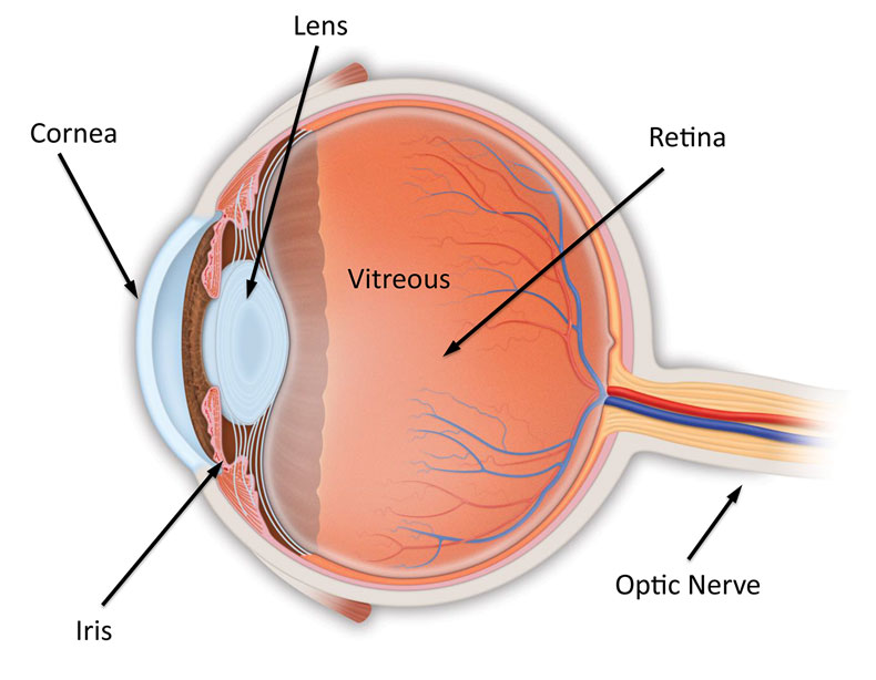
The normal human retina fundus photo shows the optic nerve (right), blood vessels and the position of the fovea (center).
![Figure 1. [The normal human retina fundus]. - Webvision - NCBI](http://webvision.org.es/wp-content/uploads/2018/01/Myopia-Fig1.jpg)
The Science Behind Myopia. Brittany J. Carr and William K. Stell - Webvision
![Figure 1. [The normal human retina fundus]. - Webvision - NCBI](https://www.pnas.org/cms/10.1073/pnas.2307380120/asset/9de33f2a-4bb0-4081-926d-bdb80222d13d/assets/images/large/pnas.2307380120fig01.jpg)
Cellular migration into a subretinal honeycomb-shaped prosthesis for high-resolution prosthetic vision
![Figure 1. [The normal human retina fundus]. - Webvision - NCBI](https://www.mdpi.com/symmetry/symmetry-15-01631/article_deploy/html/images/symmetry-15-01631-g001.png)
Symmetry, Free Full-Text
![Figure 1. [The normal human retina fundus]. - Webvision - NCBI](https://media.springernature.com/m685/springer-static/image/art%3A10.1038%2Fs41598-021-04323-3/MediaObjects/41598_2021_4323_Fig3_HTML.jpg)
Asymmetry between right and left fundus images identified using convolutional neural networks
![Figure 1. [The normal human retina fundus]. - Webvision - NCBI](http://webvision.med.utah.edu/imageswv/glaucretina.jpeg)
Simple Anatomy of the Retina : 네이버 블로그
![Figure 1. [The normal human retina fundus]. - Webvision - NCBI](https://media.springernature.com/lw685/springer-static/image/chp%3A10.1007%2F978-3-030-25886-3_22/MediaObjects/436773_1_En_22_Fig1_HTML.png)
Image Analysis for Ophthalmology: Segmentation and Quantification of Retinal Vascular Systems
![Figure 1. [The normal human retina fundus]. - Webvision - NCBI](http://webvision.org.es/wp-content/uploads/2017/01/Fig01.png)
Retinal Degeneration, Remodeling and Plasticity. Bryan William Jones, Robert E. Marc and Rebecca L. Pfeiffer - Webvision
![Figure 1. [The normal human retina fundus]. - Webvision - NCBI](https://media.springernature.com/lw685/springer-static/image/art%3A10.1186%2Fs13024-023-00655-y/MediaObjects/13024_2023_655_Fig1_HTML.png)
Retinal ganglion cell repopulation for vision restoration in optic neuropathy: a roadmap from the RReSTORe Consortium, Molecular Neurodegeneration
![Figure 1. [The normal human retina fundus]. - Webvision - NCBI](https://onlinelibrary.wiley.com/cms/asset/cbf0fdbf-c90f-440d-8bff-0bb0ec3ba7db/aos15713-fig-0002-m.jpg)
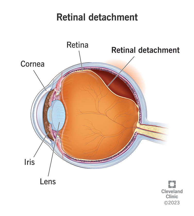
Retinal damage extends beyond the border of the detached retina in fovea‐on retinal detachment - Ng - Acta Ophthalmologica - Wiley Online Library
![Figure 1. [The normal human retina fundus]. - Webvision - NCBI](https://www.ncbi.nlm.nih.gov/books/NBK554060/bin/466648_1_En_8_Fig1_HTML.jpg)
Fig. 8.1, [Cross-sectional OCT image of human retina with the corresponding cellular structures]. - High Resolution Imaging in Microscopy and Ophthalmology - NCBI Bookshelf
![Figure 1. [The normal human retina fundus]. - Webvision - NCBI](https://www.ncbi.nlm.nih.gov/books/NBK11553/bin/clinicalergf24.jpg)
Figure 24, [Fundus photo and bright-flash ERG of patient with retinoschisis.]. - Webvision - NCBI Bookshelf
![Figure 1. [The normal human retina fundus]. - Webvision - NCBI](http://webvision.med.utah.edu/wp-content/uploads/2015/10/ArdenFig4new.jpg)
Diabetic Retinopathy and A Novel Treatment Based On The Biophysics Of Rod Photoreceptors And Dark Adaptation by Geoffrey. B. Arden and David J. Ramsey – Webvision
de
por adulto (o preço varia de acordo com o tamanho do grupo)