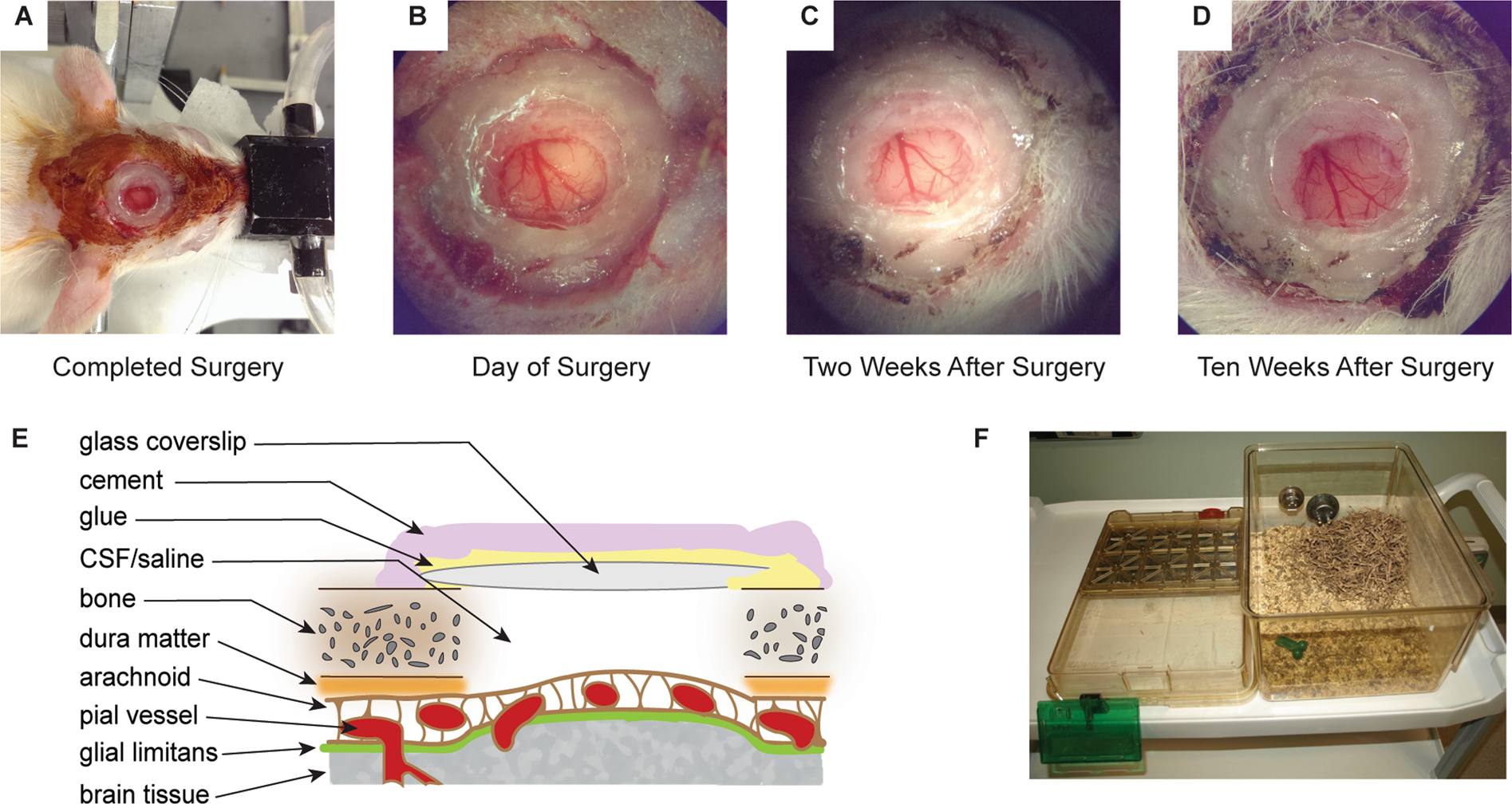
Figure 1 from Brain surface temperature under a craniotomy.
Por um escritor misterioso
Descrição
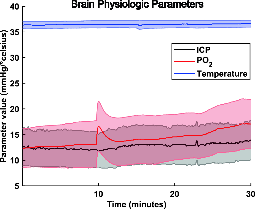
Fig. 1. Rapid cooling of the brain surface in an in vivo mouse preparation. A: schematic representation of a cranial window during recording of temperature and single-cell activity in the anesthetized mouse. The main potential routes of heat transfer are indicated. B: brain surface temperature measured with the thermocouple during replacement of the artificial cerebrospinal fluid (ACSF) with fresh ACSF warmed to 38°C. ACSF was replaced twice, indicated by the arrowheads. - "Brain surface temperature under a craniotomy."

Figure 1 from Brain surface temperature under a craniotomy.

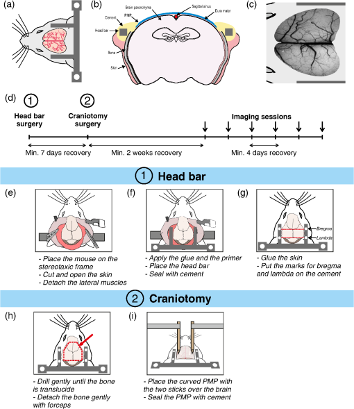
Data collection and craniotomy. Left: The infrared camera setup is

Regional temperature and quantitative cerebral blood flow responses to cortical spreading depolarization in the rat - Chunyan Li, Raj K Narayan, Ping Wang, Jed A Hartings, 2017
Brain Sciences, Free Full-Text

Temporal/Subtemporal Craniotomy

Therapeutic Hypothermia And Neuroprotection
Refinement of a chronic cranial window implant in the rat for longitudinal in vivo two–photon fluorescence microscopy of neurovascular function

Microprism-based two-photon imaging of the lateral cortex of the mouse inferior colliculus reveals novel organizational principles of the auditory midbrain
Cranial window for longitudinal and multimodal imaging of the whole mouse cortex

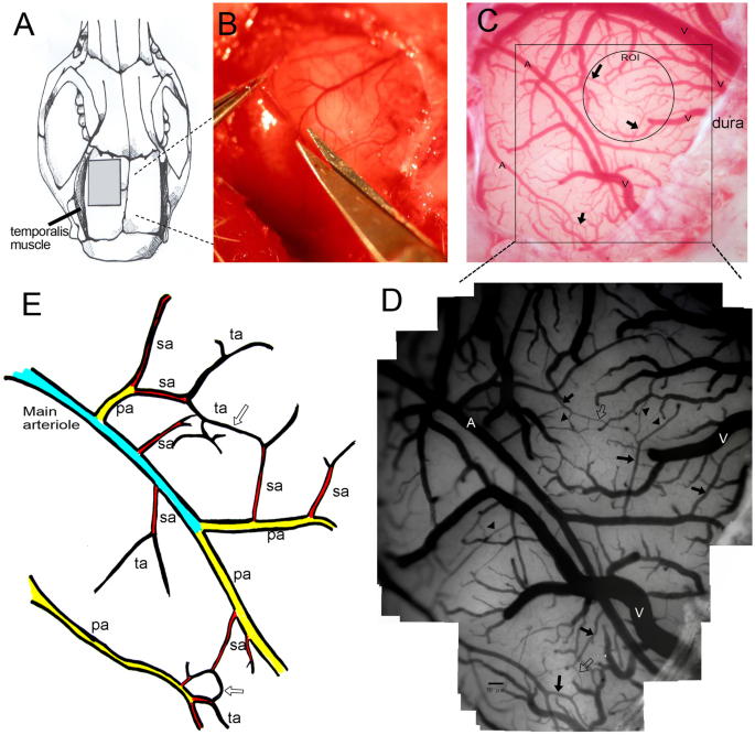
Surgical procedure for the MCA ligation. (A) After retraction of the

Altered Cortical Trigeminal Fields Excitability by Spreading Depolarization Revealed with in Vivo Functional Ultrasound Imaging Combined with Electrophysiology
The cellular coding of temperature in the mammalian cortex

Craniotomy, Expert Surgeon
Recording of pig neuronal activity in the comparative context of the awake human brain
Impaired microcirculation after subarachnoid hemorrhage in an in vivo animal model
de
por adulto (o preço varia de acordo com o tamanho do grupo)